His Check Kit (HICA)
His Check Kit (HICA)

Product Details
Product Details
Product Specification
| Host | Human |
| Stability & Storage | -80℃ |
Background
This kit uses a homogeneous immunochemiluminescence assay (HICA) based on the double-antibody sandwich principle to detect cytokine concentrations, with simple operation and no need for washing.
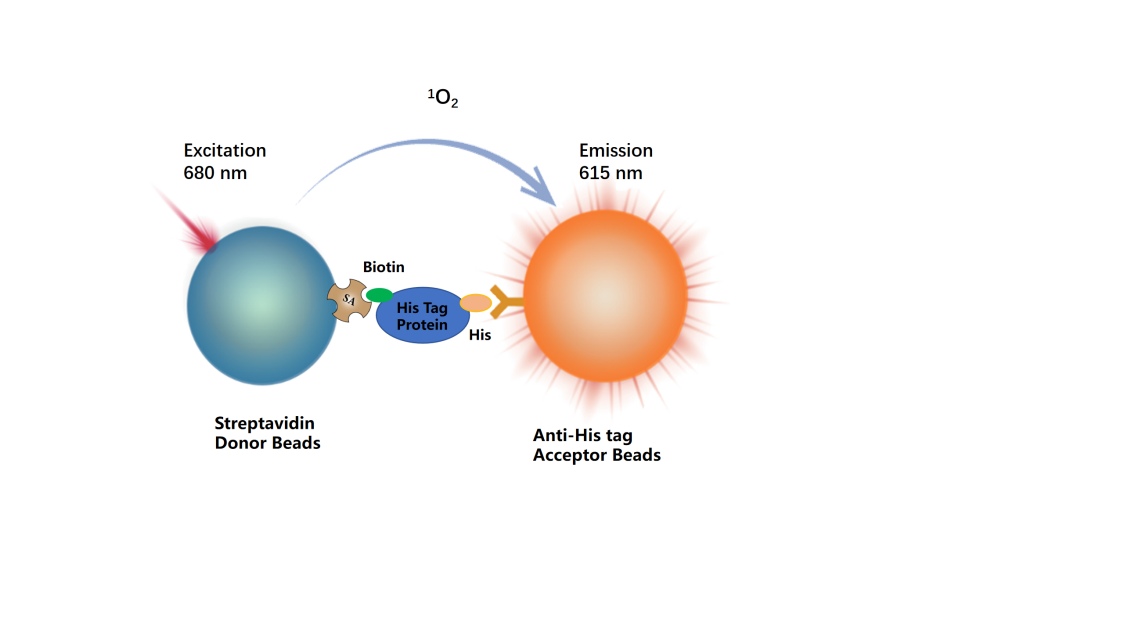
The detection system includes two types of microspheres: receptor microspheres coupled with antibodies againstHis Tag, and donor microspheres coupled with streptavidin. During the reaction,His Tag Proteinbinds totwomicrospheres, forming an immune complex that brings the two microspheres into close proximity. When the distance between the donor and receptor microspheres is less than 200 nm, singlet oxygen generated upon light excitation can be transferred to the receptor microsphere, triggering chemiluminescence;When other His-tagged proteins are present, they compete with His Tag Protein for bindingAnti-His Tag Acceptor Beads, causing the donor microbead and the receptor microbead tomove apart,resulting in aweakened signal, where the degree of signal attenuation is proportional to the concentration of the His-tag protein.
By measuring the intensity of the chemiluminescent signal, quantitative analysis of the target protein can be achieved. This method is characterized by its simple operation, rapid reaction, and high sensitivity.
Components
Component |
Concentration |
100T |
500T |
2500T |
10000T |
Storage Temperature |
His TagProtein |
20× |
25μL |
125μL |
625μL |
2500μL |
-80℃ |
Anti-His Tag Acceptor Beads |
200× |
5μL |
13μL |
65μL |
260μL |
2-8℃ |
Streptavidin Donor Beads |
125× |
5μL |
20μL |
100μL |
400μL |
2-8℃ |
Detection buffer |
1 × |
3ml |
15mL |
75mL |
300mL |
2-8℃ |
Protocol
1,Reagent Preparation
1.1Before use, prepare all reagents.Return to room temperature. The 384-well plate reaction system is 20.muL (reagent volumes for the reaction system are shown in the table). Calculate the required reaction volume before preparation and prepare accordingly. The following formulation is for reference only, using 500 reactions as an example.
Table 2.Reagent Preparation
Reagent name |
Configuration |
μL per test well |
His TagProtein |
take125 μL20 × His TagProteinThe stock solution is added to 2.375mL 1× Detection buffer dilutionExplanationTo 1×, mix well and set aside. |
5 μL |
Anti-His Tag Acceptor Beads |
take13μL200 ×Anti-His Tag Acceptor Beads, added to 2.4mL 1× Detection buffer dilutionExplanationTo 1×, mix well and set aside. |
5 μL |
Streptavidin Donor Beads |
takeTwentyμL125timesStreptavidin Donor Beads, add to 2.480mL 1× Detection buffer dilutediluteto 1×, mix well and set aside. |
5μL |
1.2sample to be testedGradient Dilution
TakingHis TagProtein 1as an example, the diluent is 1× Detection buffer,to reduce the influence of matrix effect interference, it is recommended to use a solution with the same matrix as the sample to be tested for dilution; andthe concentration of the sample to be tested should be adjusted according to its actual concentration.
Table 3.Protein to be testedHis TagProtein 1Gradient Dilution(adjust according to the actual situation)
His TagProtein 1Preparation Concentration (nM) |
Preparation Method |
|
① |
4000 |
4μL50μMHis TagProtein 1stock solution+56μL 1× Detection buffer |
② |
800 |
10μL①+40μL 1× Detection buffer |
③ |
160 |
10μL②+40μL 1× Detection buffer |
④ |
32 |
10μL③+40μL 1× Detection buffer |
⑤ |
6.4 |
10μL④+40μL 1× Detection buffer |
⑥ |
1.28 |
10μL⑤+40μL 1× Detection buffer |
⑦ |
0.256 |
10μL⑥+40μL 1× Detection buffer |
⑧ |
0.0512 |
10μL⑦+40μL 1× Detection buffer |
⑨ |
0.01024 |
10μL⑧+40μL 1× Detection buffer |
Blank |
0 |
40μL 1× Detection buffer |
Note: Different His-tagged proteins may exhibit competitive binding due to differences in molecular weight and the number of His tags.Anti-His Tag Acceptor BeadsThe capabilities also differ, so before the formal experiment, it is necessary to adjust the cell count or the concentration of the His Tag Protein to be measured to ensure a complete S-curve can be obtained.
2,Sample additionProcess
Detection process |
Detection process 1 (37℃ rapid detection) |
Detection process 2 (room temperature detection) |
Step one: |
Add 5 µL of sample in sequence,5 μLAnti-His Tag Acceptor BeadsAdd 5 µL of sample in sequence,5 μL |
Anti-His Tag Acceptor BeadsIncubationShake and mix at 400 r/min for 1 minute, incubate at 37°C for 15 minutesShake and mix at 400 r/min for 1 minute, incubate at room temperature for 60 minutes |
Step two: |
Add |
5 |
µLHis Tag |
Protein,5µL Donor Beads,5µL Donor Beads,Avoid light/green light |
Add5µL His TagProtein,5µL Donor Beads,Avoid light/green light |
Incubation |
Shake and mix at 400 r/min for 1 minute, incubate at 37°C for 10 minutes,Protect from light/green light |
Shake and mix at 400 r/min for 1 minute, incubate at room temperature for 30 minutes,Protect from light/green light |
Read the results |
Instrument reading,Protect from light/green light |
Instrument reading,Protect from light/green light |
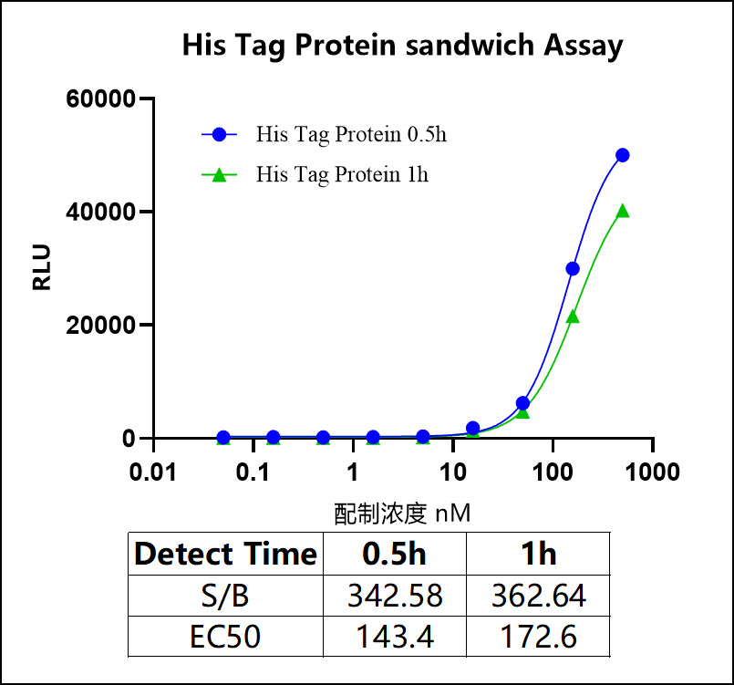
[Data display]
The following data cannot replace the data obtained in experiments; it is only for demonstration purposes, and the results mayvary depending on the plate readerandthe instrument.
1. His Tag Protein sandwich assay
2. Test results of different His Tag proteins
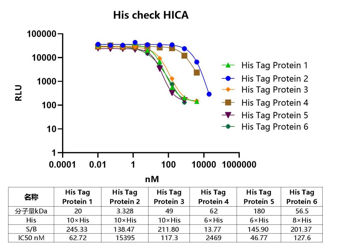
Note: Microplates are recommended(384-well plates, white, shallow wells)